Nuclear Graphite
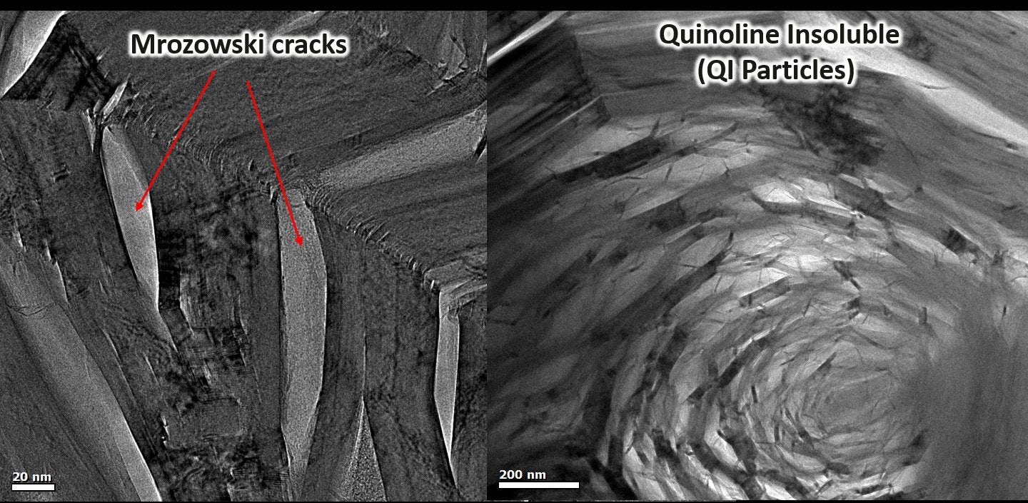
Next-generation nuclear reactors (Gen IV) will be high-temperature, gas-cooled reactors with graphite used as a core and moderator material. These reactors involve high operating temperatures as well as very high neutron dose rates, placing increased emphasis on strength, creep, and creep-fatigue behavior of the materials used. There have been no thorough studies addressing the lack of data on the creep mechanisms of graphite (i.e., dislocation movement or lack thereof), the lack of experimental data for irradiation induced dimensional changes, or the inter-relationship between these important mechanisms. Our research focuses on these fundamental issues and provide experimental data to validate or invalidate current theories. The lack of consensus on current theories can be associated with the difficulty in monitoring the dynamic atomic response of graphite subjected to irradiation. In addition, nuclear graphite has a complex microstructure composed of regions of filler, binder and turbostratic graphite phase all with varying responses to irradiation. Within the binder phase additional microstructure features may be found such as: quinoline insoluble (QI) particles, micropores, and lenticular cracks nanometers to hundreds of nanometers in length known as Mrozowski cracks.
Monitoring of the atomic level response of graphite in nuclear reactors remains impossible; however, in situ transmission electron microscopy (TEM) provides a method to monitor the dynamic atomic response of graphite during high-temperature irradiation. Specifically, in the case of graphite, high energy electrons which are used to form an image in the TEM induce irradiation damage which may be arguably comparable to irradiation from neutrons found in nuclear reactors. As such, this allows us to monitor various atomic mechanisms ‘live time’ in an environment comparable to that of a nuclear reactor.
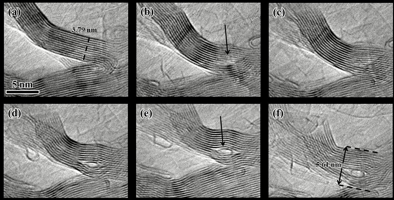
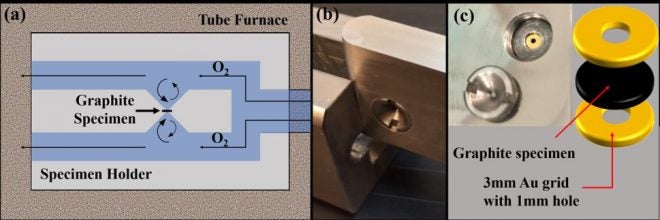
Current high-temperature electron irradiation studies, with the use of a TEM, has been shown to be a suitable method for simulating the irradiating environment graphite experiences inside a nuclear reactor. These studies have discovered additional atomic mechanisms (via the formation of carbon nanostructures) resulting in localized deformation to the graphite structure. These results provide additional explanations for changes in the properties of nuclear graphite subjected to high-temperature irradiation. In addition, we have developed novel TEM sample preparation techniques for graphite via oxidation. TEM specimens prepared via oxidation do not contain irradiation-induced artifacts which may result from conventual techniques such as ion-milling. As such, any experimental observations on oxidized specimens are unambiguous and furthermore, these specimens may be used as baseline specimens.