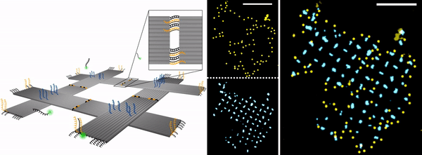
August 18, 2017 – Christopher Green, a PhD student in the Nanoscale Materials and Devices group, recently reported a new technique for characterizing DNA origami arrays with optical super-resolution microscopy. DNA origami arrays have wide-ranging potential applications, including electronic device templating, metamaterials engineering, and biomarker sensor development for disease detection. Studies of DNA origami arrays have previously relied on topological characterization techniques, such as atomic force microscopy (AFM), for array imaging. As an alternative to AFM, the team developed crystal-PAINT, a two-step super-resolution methodology for characterizing the periodic structure and quality of two-dimensional DNA origami arrays. Crystal-PAINT is the first demonstration of defect metrology for DNA origami arrays, enabling the detection of point defects, grain boundaries, and lattice misalignment in DNA arrays. The work, entitled “Metrology of DNA arrays by super-resolution microscopy,” was published recently in Nanoscale (DOI: 10.1039/c7nr00928c) and was supported by the National Science Foundation through the Scalable NanoManufacturing grant (CMMI-1344915).