Jackson Thieme, Mason Marosvari, and Dr. Laxman Mainal
Introduction
Eye lens fiber cell plasma membranes are loaded with extremely high cholesterol (Chol) content. In human lens fiber cell membrane, the Chol to phospholipid (Chol/PL) molar ratio ranges from 1:1 to 2:1 in the cortex, as high as 3:1 to 4:1 in the lens nucleus. There are four major lipids that builds fiber-cell plasma membrane of eye lens i.e., phosphatidylcholine (PC), sphingomyelin (SM), phosphatidylethanolamine (PE) and phosphatidylserine (PS). The PC is more dominant in younger animals like porcine and bovine whereas SM is more dominant in human eye lens membrane. In the proposed research the lipid membrane chosen for the study is PC.
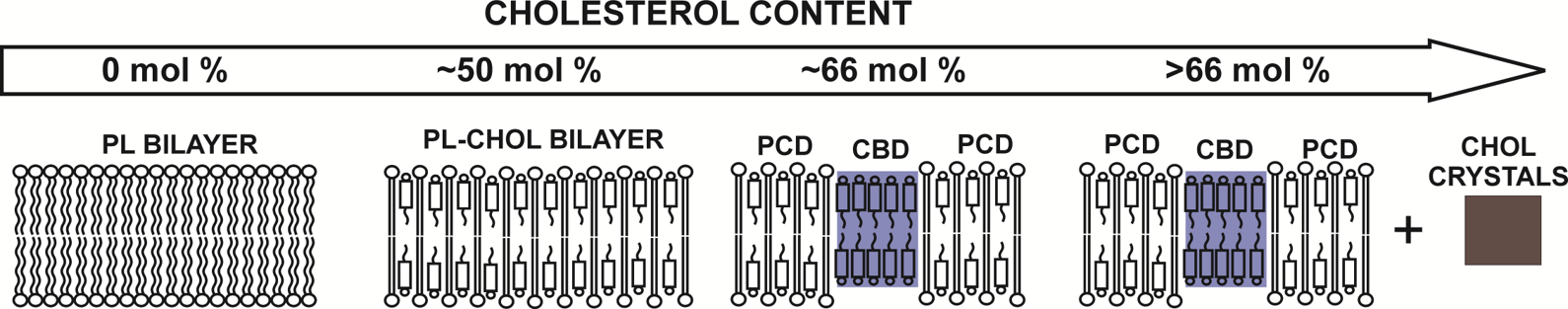
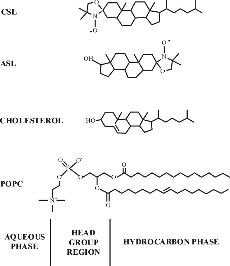
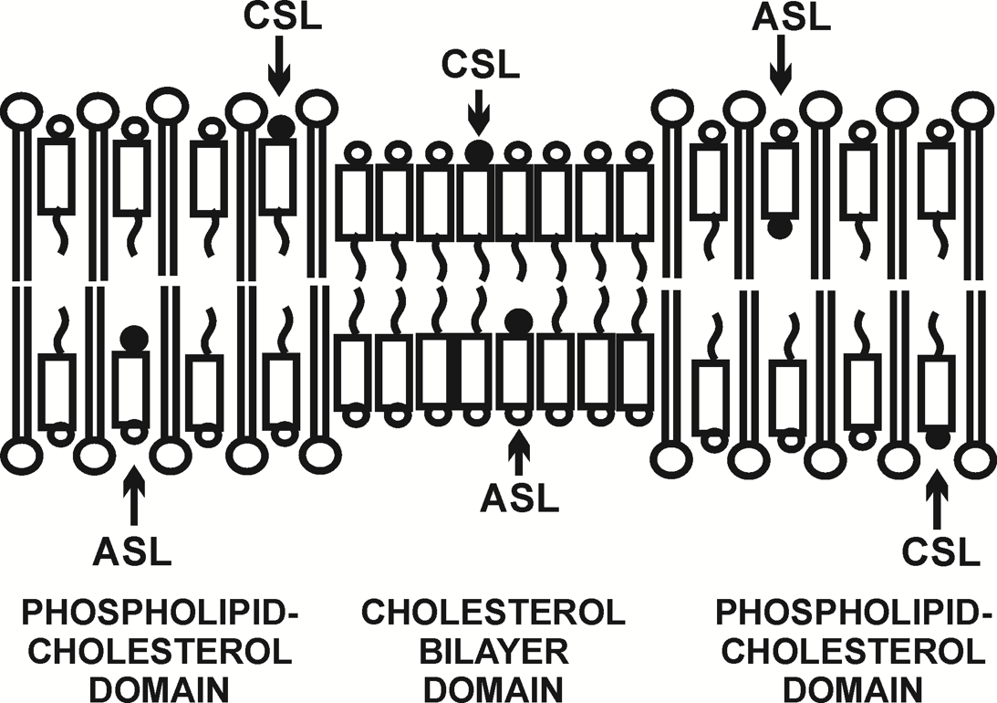
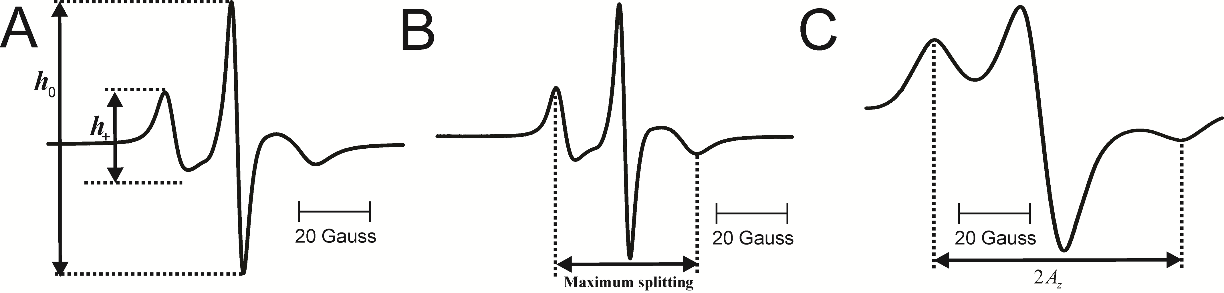
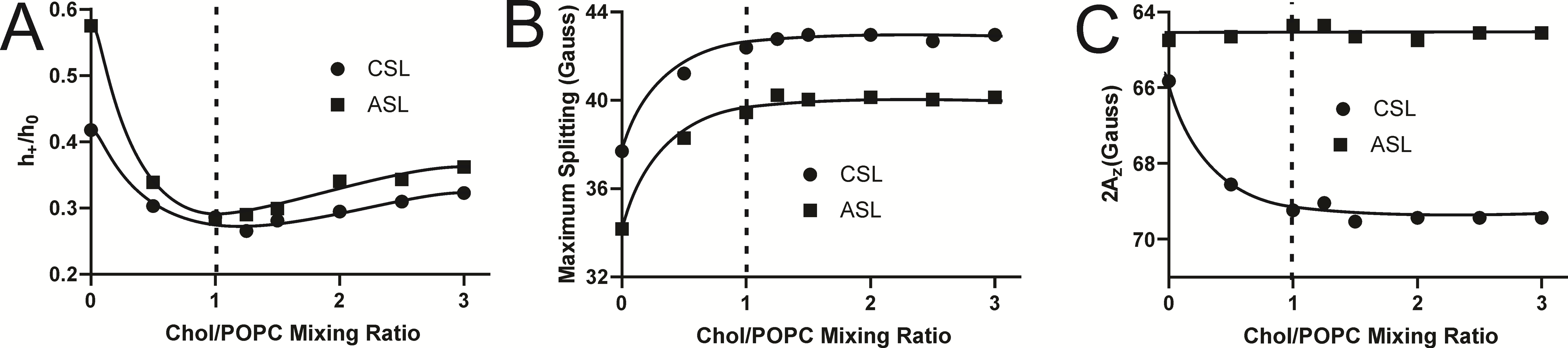
Electron paramagnetic resonance spin-labeling methods were used to investigate the physical properties of Chol/POPC (1-palmitoyl-2-oleoylphosphatidylcholine) membranes. The Chol/POPC mixing ratio was changed from 0 to 3. The membrane samples were prepared using the rapid solvent exchange method to preserve the compositional homogeneity throughout the membrane suspension. The high Chol content not only saturates the PL bilayer forming phospholipid cholesterol domain (PCD) but also leads to the formation of cholesterol bilayer domains (CBDs) as shown in Fig. 1. Recently, it was shown that CBDs start to form at Chol/POPC mixing ratio of 1 [1]. Here we have investigated the physical properties of the membranes with increasing Chol content using cholesterol analogue spin labels CSL and ASL. Physical properties include maximum splitting, mobility parameter, and hydrophobicity.
EPR Spin-Labeling Methods
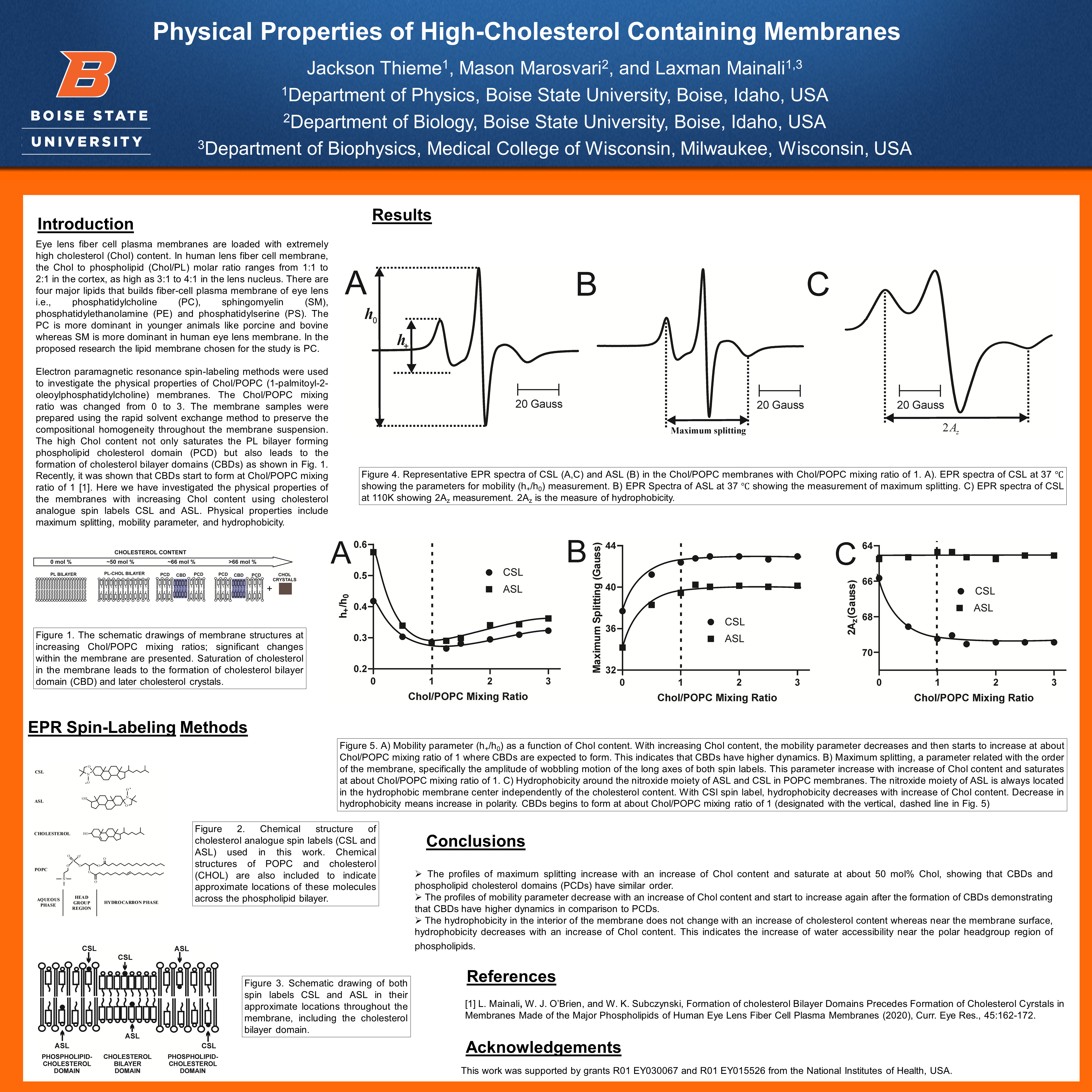
Results
Conclusions
- The profiles of maximum splitting increase with an increase of Chol content and saturate at about 50 mol% Chol, showing that CBDs and phospholipid cholesterol domains (PCDs) have similar order.
- The profiles of mobility parameter decrease with an increase of Chol content and start to increase again after the formation of CBDs demonstrating that CBDs have higher dynamics in comparison to PCDs.
- The hydrophobicity in the interior of the membrane does not change with an increase of cholesterol content whereas near the membrane surface, hydrophobicity decreases with an increase of Chol content. This indicates the increase of water accessibility near the polar headgroup region of phospholipids.
References
[1] L. Mainali, W. J. O’Brien, and W. K. Subczynski, Formation of cholesterol Bilayer Domains Precedes Formation of Cholesterol Cyrstals in Membranes Made of the Major Phospholipids of Human Eye Lens Fiber Cell Plasma Membranes (2020), Curr. Eye Res., 45:162-172.
Acknowledgements
This work was supported by grants R01 EY030067 and R01 EY015526 from the National Institutes of Health, USA.
Additional Information
For questions or comments about this research, contact Jackson Thieme at jacksonthieme@u.boisestate.edu.