27. Exploring Antibiotic Alternatives by Inhibiting Bacterial Quorum Sensing
Aliona Chernish, Cole Shaffer, Dr. Shibani Basu, and Dr. Rajesh Nagarajan
Select to view full poster image
Introduction
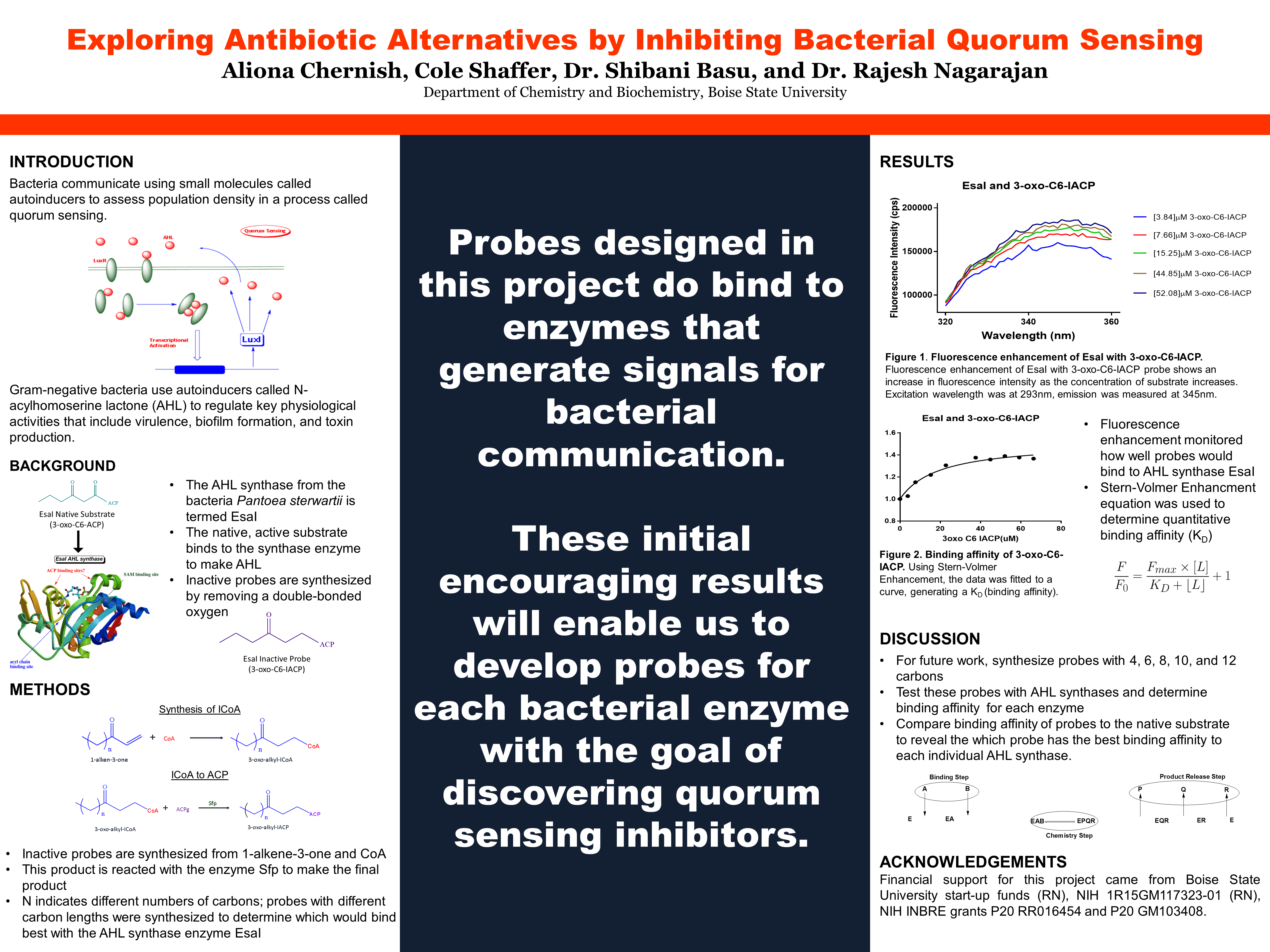
Bacteria communicate using small molecules called autoinducers to assess population density in a process called quorum sensing.
This image shows the basic mechanism of bacterial communication. A signal molecule attaches to a receptor on the cell surface. The signal/receptor complex then travels to the DNA and transcribes an enzyme that makes more signal molecules.
Gram-negative bacteria use autoinducers called N-acylhomoserine lactone (AHL) to regulate key physiological activities that include virulence, biofilm formation, and toxin production.
Background
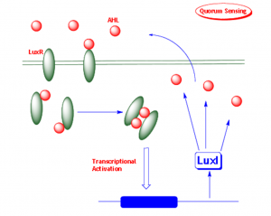
The AHL synthase from the bacteria Pantoea sterwartii is termed EsaI The native, active substrate binds to the synthase enzyme to make AHL Inactive probes are synthesized by removing a double-bonded oxygen
Methods
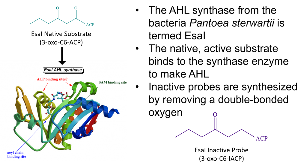
This image shows the reaction equation to make the lab synthesized inactive signal molecule. A 1-alkene-3-one plus a CoA molecule makes a 3-oxo-alkyl-ICoA. As well as the next step to make the inactive signal molecule. The 3-oxo-alkyl-ICoA plus the protein ACPg make -oxo-alkyl-IACP. The enzyme Sfp is used to catalyze this reaction.
Inactive probes are synthesized from 1-alkene-3-one and CoA
This product is reacted with the enzyme Sfp to make the final product
N indicates different numbers of carbons; probes with different carbon lengths were synthesized to determine which would bind best with the AHL synthase enzyme EsaI
Results
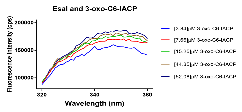
Figure 1. Fluorescence enhancement of EsaI with 3-oxo-C6-IACP. Fluorescence enhancement of EsaI with 3-oxo-C6-IACP probe shows an increase in fluorescence intensity as the concentration of substrate increases. Excitation wavelength was at 293nm, emission was measured at 345nm.Figure 2. Binding affinity of 3-oxo-C6-IACP. Using Stern-Volmer Enhancement, the data was fitted to a curve, generating a KD (binding affinity).
Fluorescence enhancement monitored how well probes would bind to AHL synthase EsaI
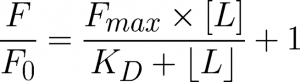
Stern-Volmer Enhancment equation was used to determine quantitative binding affinity (KD)
This is the formula from the Stern-Volmer Enhancement equation. This equation is used to find the binding affinity (KD) of an enzyme to its substrate.
Discussion
For future work, synthesize probes with 4, 6, 8, 10, and 12 carbons
Test these probes with AHL synthases and determine binding affinity for each enzyme
Compare binding affinity of probes to the native substrate to reveal the which probe has the best binding affinity to each individual AHL synthase.
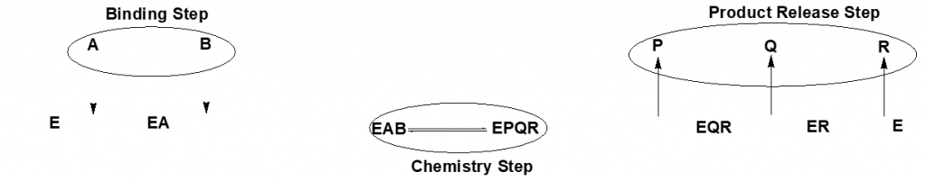
This graphic shows in what order molecules bind to and enzyme and release from it. The enzyme E first forms a complex with molecule A to form EA. Then molecule B attaches to form EAB. A reaction occurs to turn A and B into P, Q, and R. P releases from the enzyme first, leaving the complex EQR. Then Q releases, then R, leaving just the enzyme E.
Acknowledgements
Financial support for this project came from Boise State University start-up funds (RN), NIH 1R15GM117323-01 (RN), NIH INBRE grants P20 RR016454 and P20 GM103408.