Topher Dayton, Eric McIndoo, Dr. Amy Bryant, Dr. Dennis Stevens, Dr. Sarah Hobdey
Background
Necrotizing fasciitis, better known as “the flesh-eating disease,” is caused by a severe infection from a number of different bacterial species but is most commonly caused by Streptococcus pyogenes, or Group A Strep (GAS). This disease is very rare, with roughly 100-11,000 cases per year in the U.S. (Stephens). However, GAS is very common throughout our environment and causes less severe infections frequently, such as strep throat. How these bacteria produce less severe infections but can then be responsible for illnesses that commonly cause amputation is poorly understood. Here, we investigated how two toxins produced by GAS, streptolysin O (SLO) and S. pyogenes NAD+ glycohydrolase (SPN), affect two different cell types, alveolar and skeletal muscle.
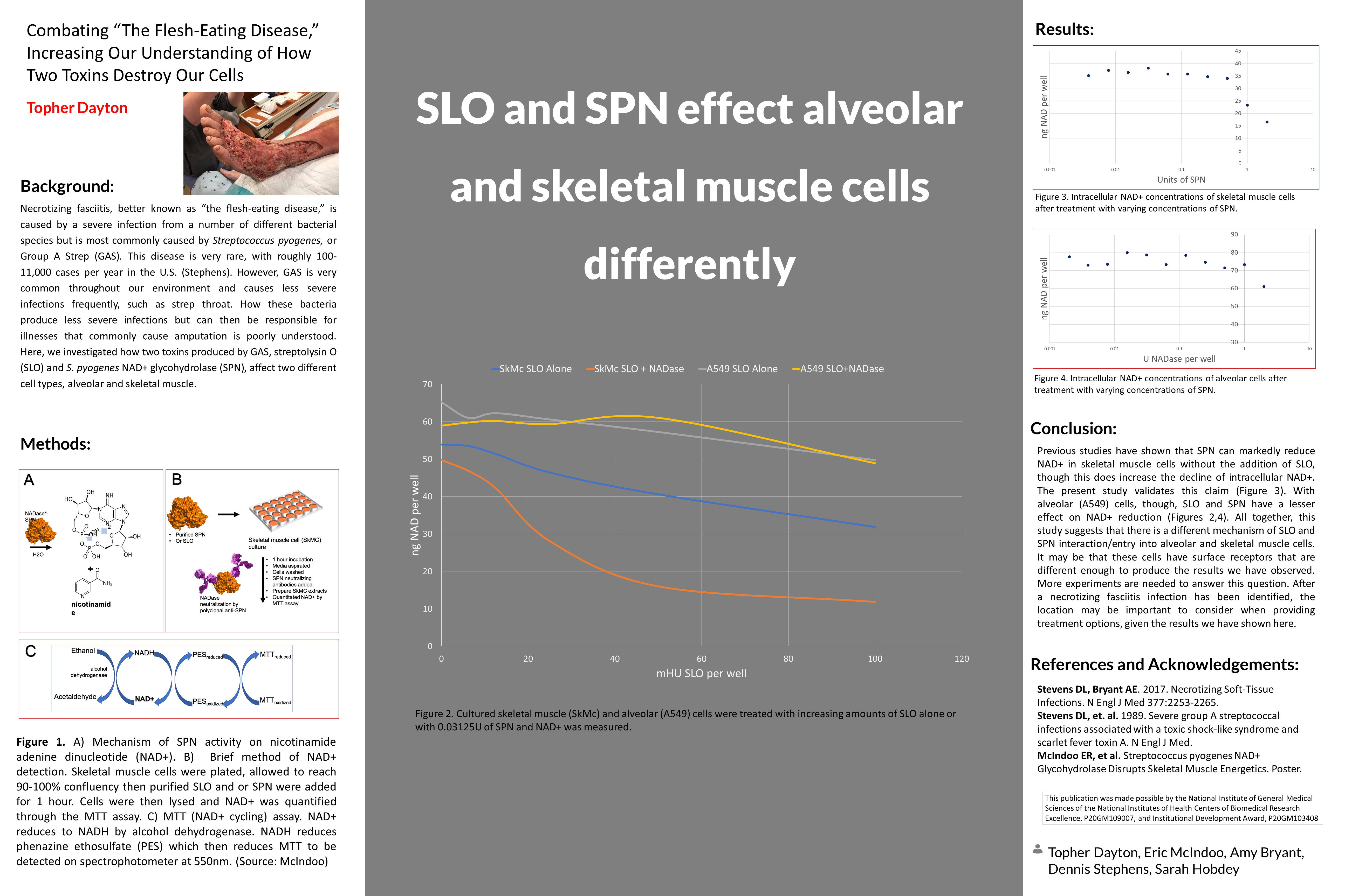
Methods
Results
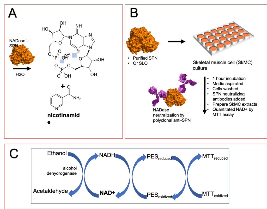
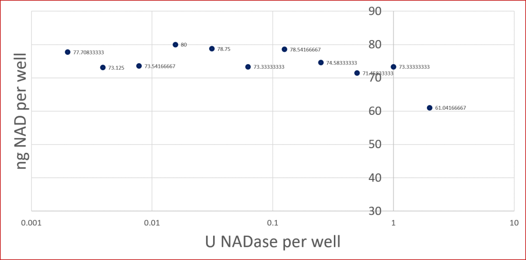
SLO and SPN effect alveolar and skeletal muscle cells differently
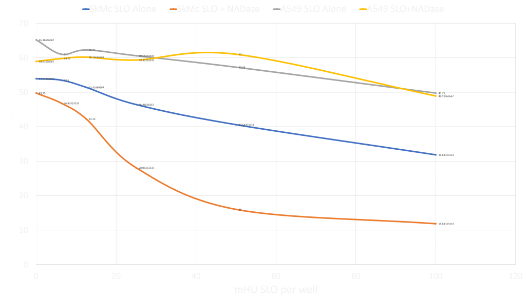
Units of Spin
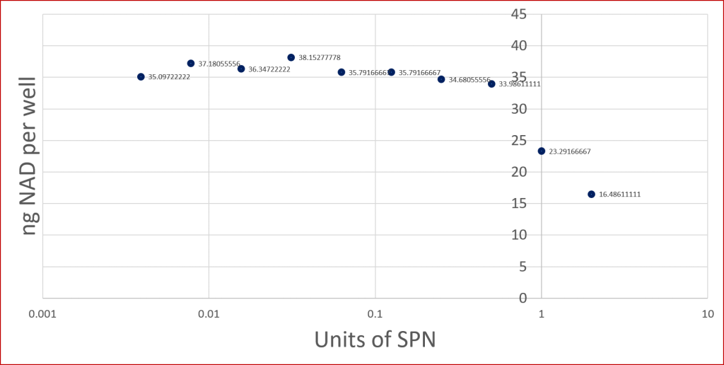
U NADase per well
Conclusion
Previous studies have shown that SPN can markedly reduce NAD+ in skeletal muscle cells without the addition of SLO, though this does increase the decline of intracellular NAD+. The present study validates this claim (Figure 3). With alveolar (A549) cells, though, SLO and SPN have a lesser effect on NAD+ reduction (Figures 2,4). All together, this study suggests that there is a different mechanism of SLO and SPN interaction/entry into alveolar and skeletal muscle cells. It may be that these cells have surface receptors that are different enough to produce the results we have observed. More experiments are needed to answer this question. After a necrotizing fasciitis infection has been identified, the location may be important to consider when providing treatment options, given the results we have shown here.
References and Acknowledgements
- Stevens DL, Bryant AE. 2017. Necrotizing Soft-Tissue Infections. N Engl J Med 377:2253-2265.
- Stevens DL, et. al. 1989. Severe group A streptococcal infections associated with a toxic shock-like syndrome and scarlet fever toxin A. N Engl J Med.
- McIndoo ER, et al. Streptococcus pyogenes NAD+ Glycohydrolase Disrupts Skeletal Muscle Energetics. Poster.
This publication was made possible by the National Institute of General Medical Sciences of the National Institutes of Health Centers of Biomedical Research Excellence, P20GM109007, and Institutional Development Award, P20GM103408
Additional Information
For questions or comments about this research, contact Tohper Dayton at topherdayton@u.boisestate.edu.