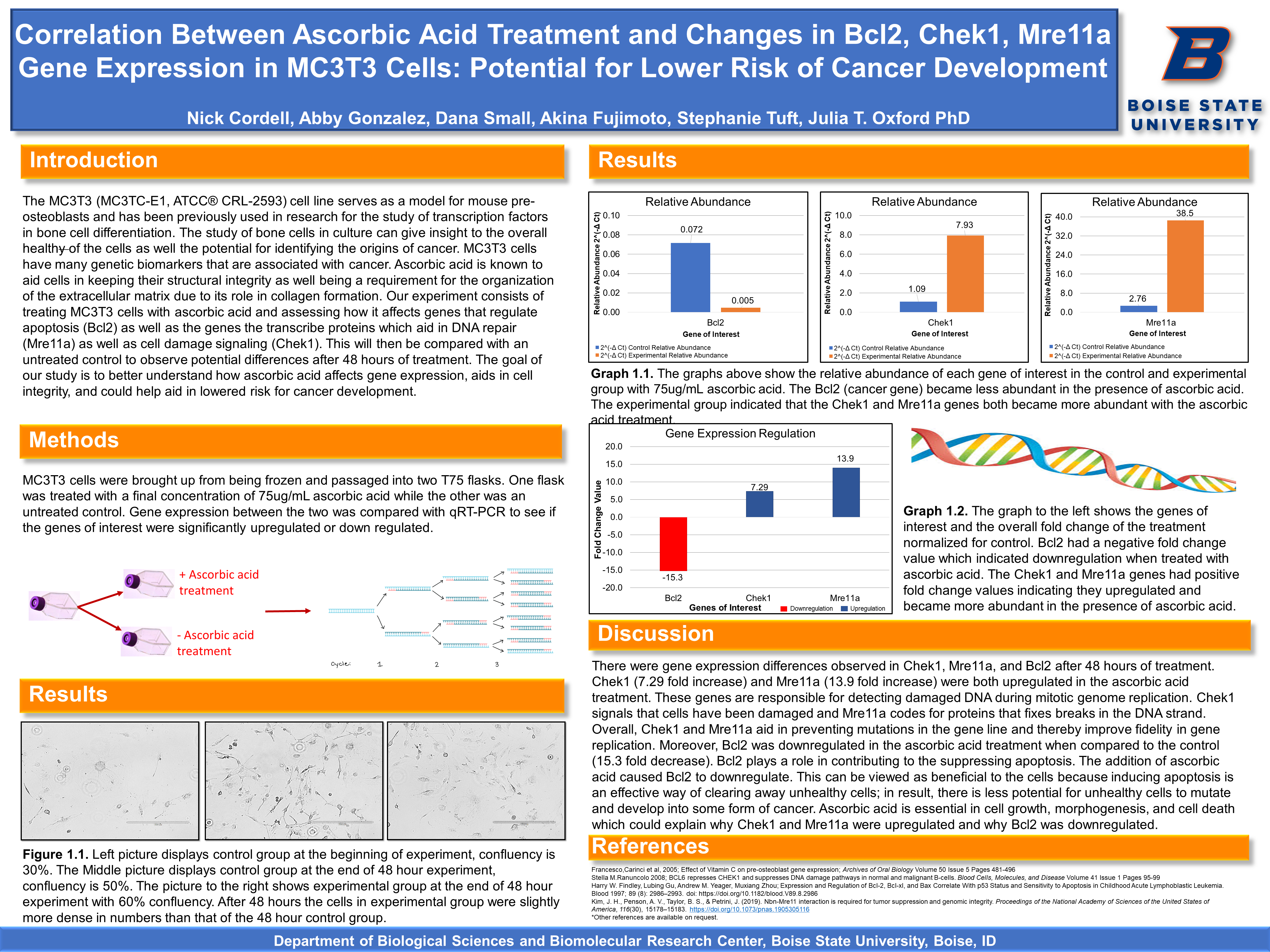
Potential for Lower Risk of Cancer Development
Nick Cordell, Abby Gonzalez, Dana Small, Akina Fujimoto, Stephanie Tuft, Dr. Julia Oxford
Introduction
The MC3T3 (MC3TC-E1, ATCC® CRL-2593) cell line serves as a model for mouse pre-osteoblasts and has been previously used in research for the study of transcription factors in bone cell differentiation. The study of bone cells in culture can give insight to the overall healthy of the cells as well the potential for identifying the origins of cancer. MC3T3 cells have many genetic biomarkers that are associated with cancer. Ascorbic acid is known to aid cells in keeping their structural integrity as well being a requirement for the organization of the extracellular matrix due to its role in collagen formation. Our experiment consists of treating MC3T3 cells with ascorbic acid and assessing how it affects genes that regulate apoptosis (Bcl2) as well as the genes the transcribe proteins which aid in DNA repair (Mre11a) as well as cell damage signaling (Chek1). This will then be compared with an untreated control to observe potential differences after 48 hours of treatment. The goal of our study is to better understand how ascorbic acid affects gene expression, aids in cell integrity, and could help aid in lowered risk for cancer development.
Methods
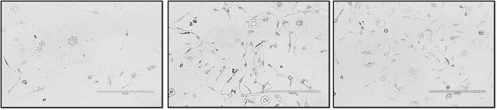
MC3T3 cells were brought up from being frozen and passaged into two T75 flasks. One flask was treated with a final concentration of 75ug/mL ascorbic acid while the other was an untreated control. Gene expression between the two was compared with qRT-PCR to see if the genes of interest were significantly upregulated or down regulated.

Results
Discussion
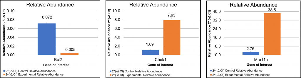
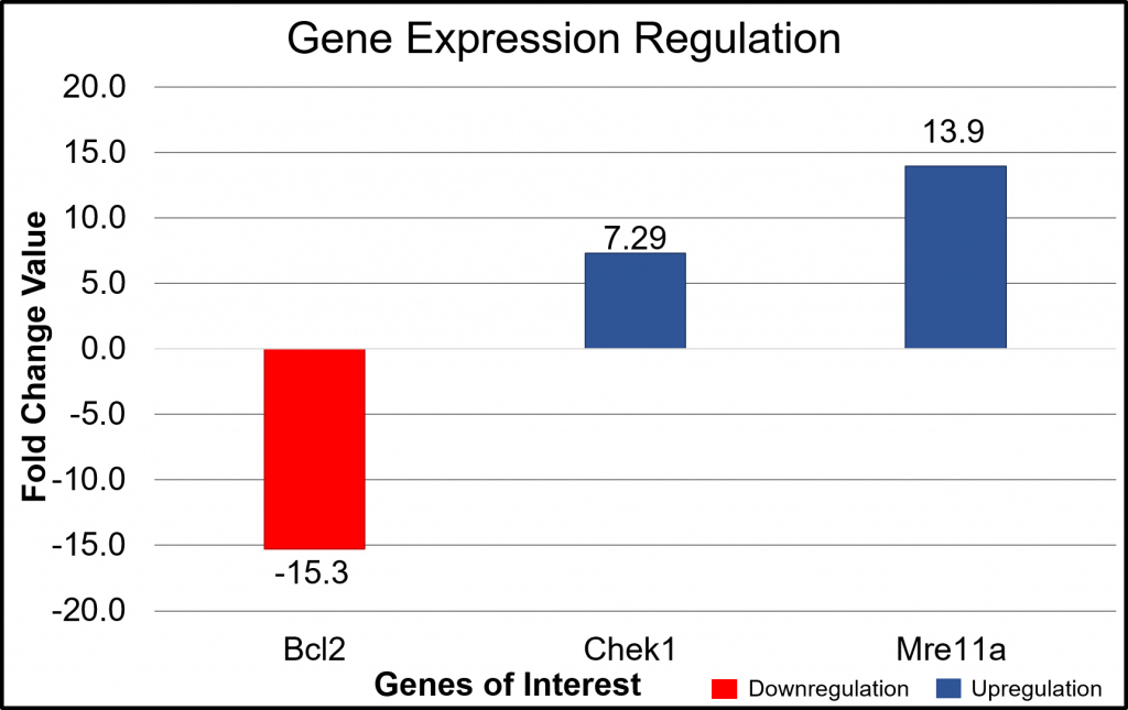
There were gene expression differences observed in Chek1, Mre11a, and Bcl2 after 48 hours of treatment. Chek1 (7.29 fold increase) and Mre11a (13.9 fold increase) were both upregulated in the ascorbic acid treatment. These genes are responsible for detecting damaged DNA during mitotic genome replication. Chek1 signals that cells have been damaged and Mre11a codes for proteins that fixes breaks in the DNA strand. Overall, Chek1 and Mre11a aid in preventing mutations in the gene line and thereby improve fidelity in gene replication. Moreover, Bcl2 was downregulated in the ascorbic acid treatment when compared to the control (15.3 fold decrease). Bcl2 plays a role in contributing to the suppressing apoptosis. The addition of ascorbic acid caused Bcl2 to downregulate. This can be viewed as beneficial to the cells because inducing apoptosis is an effective way of clearing away unhealthy cells; in result, there is less potential for unhealthy cells to mutate and develop into some form of cancer. Ascorbic acid is essential in cell growth, morphogenesis, and cell death which could explain why Chek1 and Mre11a were upregulated and why Bcl2 was downregulated.
References
- Francesco,Carinci et al, 2005; Effect of Vitamin C on pre-osteoblast gene expression; Archives of Oral Biology Volume 50 Issue 5 Pages 481-496
- Stella M.Ranuncolo 2008; BCL6 represses CHEK1 and suppresses DNA damage pathways in normal and malignant B-cells. Blood Cells, Molecules, and Disease Volume 41 Issue 1 Pages 95-99
- Harry W. Findley, Lubing Gu, Andrew M. Yeager, Muxiang Zhou; Expression and Regulation of Bcl-2, Bcl-xl, and Bax Correlate With p53 Status and Sensitivity to Apoptosis in Childhood Acute Lymphoblastic Leukemia. Blood 1997; 89 (8): 2986–2993. doi: https://doi.org/10.1182/blood.V89.8.2986
- Kim, J. H., Penson, A. V., Taylor, B. S., & Petrini, J. (2019). Nbn-Mre11 interaction is required for tumor suppression and genomic integrity. Proceedings of the National Academy of Sciences of the United States of America, 116(30), 15178–15183. https://doi.org/10.1073/pnas.1905305116
*Other references are available on request.