- About the Facility
- Faculty/Staff, Contact Information
- Instrument Capabilities – BD Influx Cell Sorter
- Cell Sorting Rates, Policies
- Applications
- Understanding Flow Cytometry
- Additional Resources
About the Facility
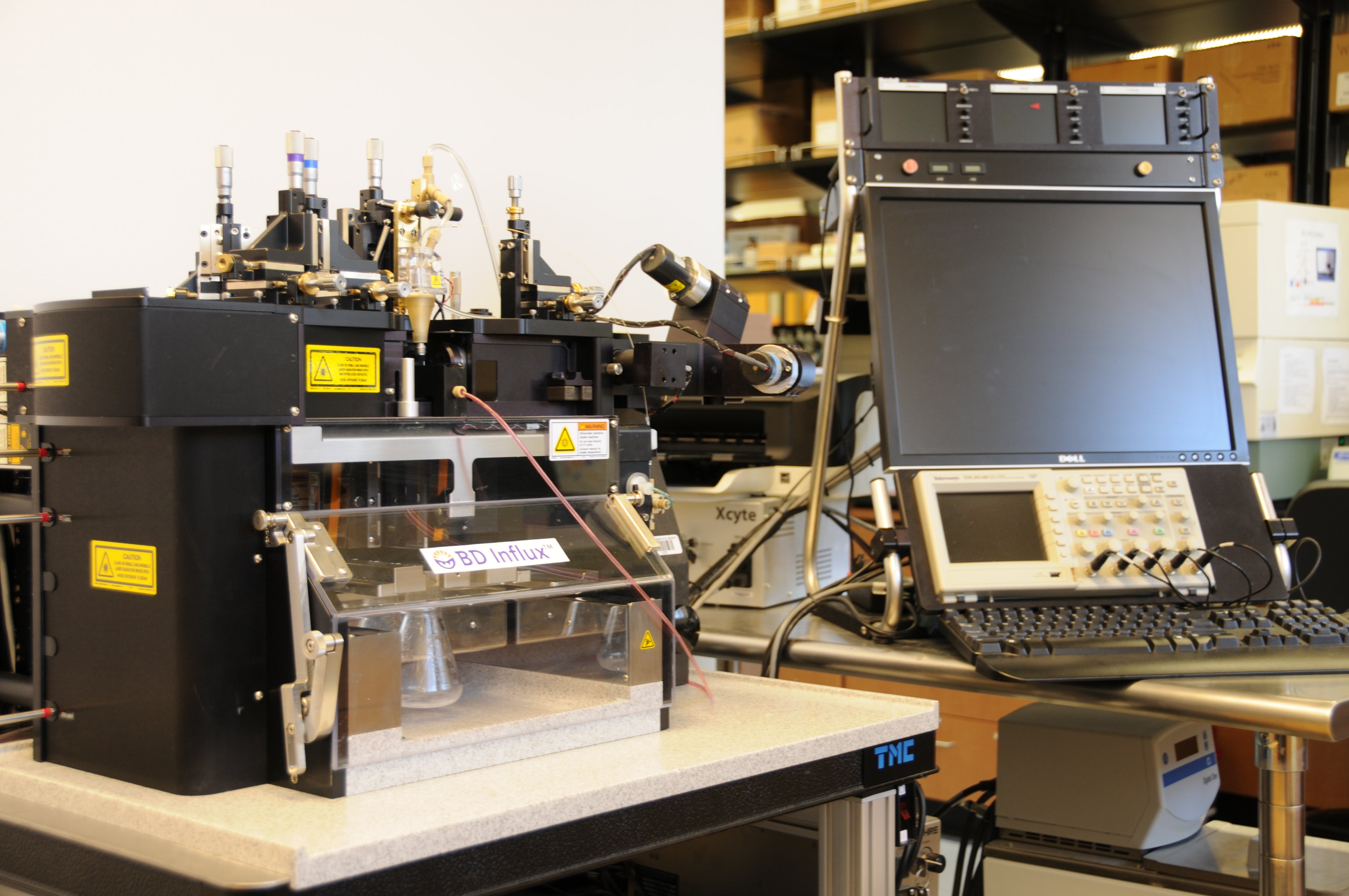
The Flow Cytometry Core Facility (FCCF) at Boise State University was created to aid researchers and graduate students in implementing flow cytometry into their experimentation. The facility’s services, housed in the Science building (Room 218), are available primarily to the Boise State University and Treasure Valley communities. The FCCF is equipped with a state of the art four-laser BD INFLUX cell sorter. This instrument supports four-way sorting, plate sorting for single cell isolation, 9-color analysis, and can operate at up to 200,000 events per second. The BD INFLUX at BSU is also equipped with a small particle detector allowing particle detection near 200 nm.
Faculty/Staff, Contact Information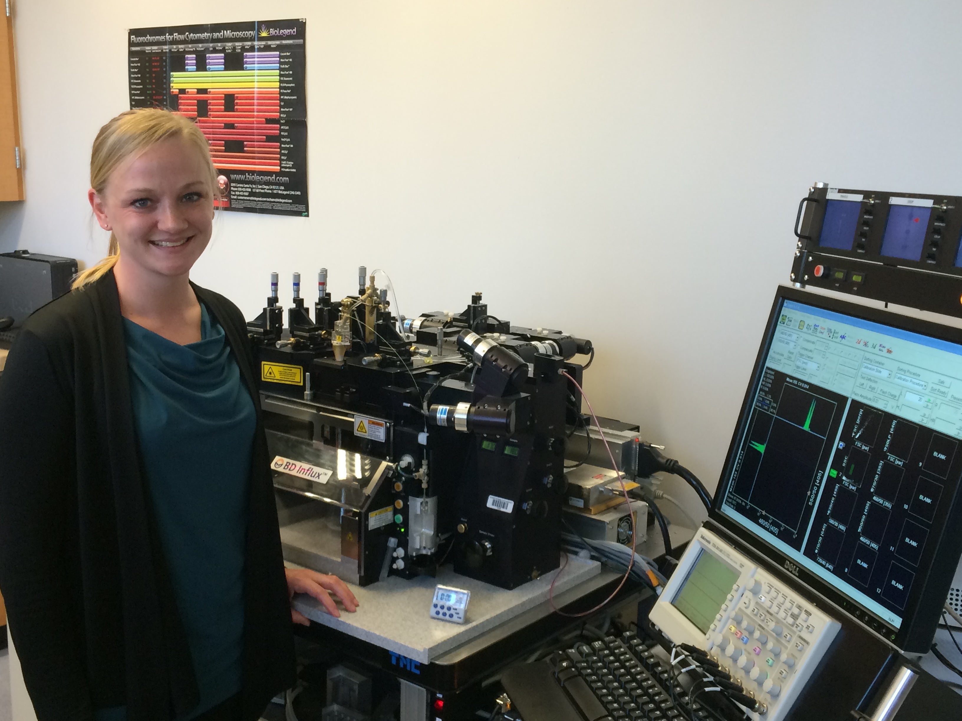
To schedule time or consultation for services, please contact Dr. Wingett.
- Dr. Denise Wingett – Director, Flow Cytometry Core Facility; Professor, Department of Biological Sciences; Director, Biomolecular Sciences Ph.D. Program; (208) 426-2921; email Dr. Wingett
Applications
There are many uses for flow cytometry, many of which are outlined in this video by Beckman Coulter. Examining, counting, and sorting cells are the primary functions of this technology. Samples are sorted primarily based on the presence of a fluorescent dye, but also size and granularity characteristics. These characteristics assist researchers in the following examples of applications of flow cytometry:
- Analyze target (Ab bound, fluorescent) versus non-target cells
- Measure DNA content of a cell sample
- Cell cycle analysis determination
- RNA vs DNA differentiation during cell cycle progression
- Distinguish between immature and mature RBC
- Evaluate cell viability
- Investigate cytokine production from activated cells
- Monitor second messengers to determine cell activation state
Understanding Flow Cytometry
Flow cytometry analyzes cells based on relative size, granularity, and fluorescence. A flow cytometer is composed of three main systems: fluidics, optics, and electronics. The fluidics system operates to inject the cell sample into the sheath fluid for delivery to the laser for interrogation. Hydrodynamic focusing of the sample within the sheath fluid allows for one cell to pass through the laser at a time. The optical system contains lasers, lenses, and filters. As a cell passes through the laser beam it will scatter light for relative size and granularity measurements. Lasers also excite fluorochromes present in the cell sample. Lenses and filters are required to direct the scattered and excited light of specific wavelengths to various detectors for collection. The electronics system converts the light signals received by the detectors to electronic signals using photodetectors (photodiodes and photomultiplier tubes). These converted light signals are displayed on a graph, such as a dot plot or histogram, that can be used to analyze, gate, or sort a selected cell population in the sample.
Additional Resources
Additional resources regarding flow cytometry can be found at the following links:
- For help selecting antibodies and designing a flow cytometry panel, visit FluoroFinder
- Beckman Coulter’s Computer Based Training: Animated Flow Cytometry Theory
- BD Bioscience Introduction to Flow Cytometry: A Learning Guide (PDF)
- BD Bioscience BD Flow Cytometry: Technical Bulletin
- Thermo Fisher Scientific Flow Cytometry Learning Center