The Mass Spectrometry Facilities mission is to provide a wide range of analytical capabilities through fee-for-service in the areas of protein, biological and small molecule research to Boise State University investigators, external academic and industrial partners. If you have any questions regarding our services or wish to know more about mass spectrometry, its capabilities, and applications, please contact us:
Xinzhu Pu, PhD
Assistant Research Professor, Mass Spectrometry and BRCF Manager
1910 University Drive
Boise, ID 83725-1511
Phone: (208)4262233
Email: shinpu@boisestate.edu
Instrumentation
The Biomolecular Research Core Facility currently has two mass spectrometers; a Bruker Daltonics maXis Quadrupole Time-of-Flight, Thermo Scientific Velos Pro Dual-Pressure Linear Ion Trap. Both instruments are located in the BRCF, room 215 in the Mathematics Building.
Bruker Daltonics maXis Quadrupole Time-of-Flight (Q-TOF) Mass Spectrometer
The Bruker Q-TOF Mass Spectrometer is a hybrid tandem mass spectrometer with outstanding performance including fast acquisition rate (up to 30 Hz for small molecules, up to 5Hz dynamic for peptides), high resolution (50,000 Full Sensitivity and Resolution), high-resolution EIC (0.5 – 1mDa on typical LC peaks), and excellent sensitivity (1 pg Reserpine >100:1 S/N RMS). This mass spectrometer is coupled with a Dionex Ultimate 3000 HPLC system and an innovative Captive electrospray source.
In combination with software tools, including Bruker Compass Data Analysis, Smartformula, ProteinScape, Mascot protein search engine, and Profile Analysis, we use this LC-MS system in small molecule identification, metabolomics analysis, and protein characterization.
Publications and presentations utilizing data from samples analyzed with the Bruker maXis Q-TOF Mass Spectrometer should include the following acknowledgment: “This research was made possible by the National Science Foundation Grant NO: 0923535.”
Thermo Scientific Velos Pro Dual-Pressure Linear Ion Trap (LIT) Mass Spectrometer
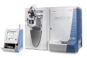
The Themo Scientific Velos Pro Linear Ion Trap Mass Spectrometer offers Trap-HCD (Higher-Energy Collisional Dissociation) combined with CID (Collision-Induced Dissociation), and PQD (Pulsed-Q Dissociation) to enhance coverage and sensitivity of proteomic analysis. An Easy nLCII nano liquid chromatographic system is coupled to the mass spectrometer through a nanoelectrospray source for protein characterization. In combination with the Thermo Proteome Discoverer 1.3, Sequest and Mascot database search engine, this LC-MS system is our work horse for routine proteomic analysis.
Publications and presentations utilizing data from samples analyzed with the LTQ Velos Mass Spectrometer should include the following acknowledgement: “This research was made possible by the INBRE Program, National Institutes of Health Grant NOs: P20 RR016454 (National Center for Research Resources) and P20 GM103408 (National Institute of General Medical Sciences), by the Idaho State Board of Education’s Higher Education Research Council (HERC) and the Biomolecular Research Core Facility at Boise State University.
Fees
We charge for every sample run, regardless of whether an expected result is obtained. The only exception is if the lack of result is caused by human errors or instrument problems in the mass spec facility.
Services
We currently provide the mass spectrometry analyses listed in this section. If you have needs which are not listed, please contact us to discuss your project with our staff. We will do our best to help you get your research done!
Mass spectrometry is a powerful tool in protein and metabolite identification and quantification. However, just like many other analytical tools, it has limitations and there are many factors that will affect the results. We strongly encourage our users to discuss their projects with our facility staff before they submit samples for analysis. We would like to discuss if mass spectrometry is suitable for your project, the best mass spec workflow for your specific needs, and how to prepare your sample for mass spec analysis. To ensure the success of analysis, samples must be prepared in a way that is compatible with mass spec. Please follow the sample preparation guide to prepare your samples for mass spec analysis.
Tryptic Digestion and Protein Identification
Protein identification by tandem MS after protease digestion is a robust tool in proteomics with high successful rates. Samples can be visible bands excised from Coomassie blue or silver-stained gels. Silver staining must be performed with mass-spec compatible reagents,e.g. Pierce Silver Stain Kit for Mass Spectrometry, catalog # 24600. We may refuse submissions stained using MS incompatible methods. We also accept samples in solution which contains purified protein or several proteins.
Please contact facility staff before you prepare your sample for in-solution digestion because some of the chemical commonly used in protein extraction are detrimental to mass spec. For complex samples, fractionation is usually required, for example, SDS-PAGE or 2D gel electrophoresis. Protein samples will be digested with protease in gel or in solution and resulted peptides will be analyzed by tandem MS. Trypsin is the most commonly used protease and is the enzyme we routinely use. Some proteins may require other proteases, such as Lys-C, Asp-N, chymotrypsin, etc. If this is the case, we can accommodate.
Guideline for Preparing gel slice/band:
Electrophoresis
- Try to use a 1 mm thick gel.
- If possible, try using precast gels and pre-made electrophoresis running buffer.
- We highly recommended to electrophorese a positive control such as BSA or some other known protein that stains to approximately the same level as your unknown protein of interest.
Band excision
- Use a clean scalpel or razor blade (you may wish to first sonicate the blade for 5 min acetonitrile or ethanol in a glass beaker) to excise protein bands/spots of interest from the stained polyacrylamide gel. Excise only the stained part(s) of the band and trim off the non-stained gel as much as possible. Gel band/slice size should be no larger than 5mm in length.
- Cut each gel slice into small pieces (~1 mm3) and transfer the pieces to a clean, sterile 1.5 mL microcentrifuge tube pre-rinsed with 50% organic solvents, such as methanol and acetonitrile.
- Also, excise and dice a gel piece from a protein-free region of the gel in parallel as a negative control, and your positive control (for example, BSA) if you ran it.
- Close the tube as soon as your bands are inside. Submit a sample to the Mass-Spec facility immediately or store gel slices at -20C for a brief period before submission.
Strategies to avoid Keratin contamination
All humans are constantly shedding skin cells so there is a large amount of keratin in the air of labs, on surfaces, and in-gel boxes, glassware etc. that have been left uncovered on benches. Large amounts of keratin in a sample can mask proteins of interest that are less abundant. Although human keratin contamination cannot be completely avoided, it is important to minimize it during each sample preparation step. Cleanliness is the key. Here are some recommendations:
- A key factor in avoiding keratin is to avoid contamination with dust particles. Start with a scrupulously clean work surface. If possible, for best dust control during sample prep work such as cutting gel bands or performing in-gel trypsin digestion, try working in a HEPA-filtered laminar-airflow hood. Wipe the inside of the hood and surface of plasticware packs with ethanol.
- DO NOT handle anything that will come into contact with your protein with your bare hands! Always wear powder-free nitrile gloves (never use latex gloves, as natural rubber contains significant amounts of keratin and other proteinaceous materials). It may be necessary to wear protective clothing during all phases of sample preparation and to cover your head.
- Try using precast gels, pre-made electrophoresis running buffer.
If casting your own gel
- Clean the gel plates well with a soapy sponge, rinsing well with ethanol (or bleach overnight).
- Handle the plates by the edges and wear gloves to avoid transfer of keratin from your hands to the gel plates.
- Before preparing solutions, wash the glassware well to remove any dust that might have accumulated during storage.
When staining gels
- Rinse the staining container well before beginning. Also, rinse the gel thoroughly at each stage. Keep the container closed to keep dust out.
When cutting gel bands
- This is the step where keratin contamination is most likely to occur. If possible, work in a laminar flow hood. Before starting, wipe down the work area and any tools that will contact the gel band with an ethanol-soaked Kimwipe.
Prepare sample for in-solution digestion
Detergents are incompatible with mass-spectrometry and must be avoided. Failure to do so will contaminate the instrument and result in costly and time-consume maintenance and serious down-time for the instruments. Therefore we require users to contact facility staff before preparing in-solution sample for submission to ensure samples are free of mass spec in=compatible reagents.
- Do not use ANY detergents in ANY step of sample-prep. This includes, but is not limited to SDS, CHAPS, NP-40, and Triton-X100 (or related).
- If detergents and other reagents that are not compatible with mass spec must be used to extract protein, precipitation procedure, for example, acetone precipitation can be used to eliminate these interfering reagents.
- Optimal sample quantity is 20 µg protein in ~20µL solution.
Protein molecular weight determination
Molecular weight determination of intact protein can be achieved by direct infusion via a syringe pump or Liquid Chromatographic (LC) – Electro Spray Ionization (ESI) on our Bruker maXis Q-TOF. A concentrated solution of desalted protein is sprayed into the instrument. Ion signals that correspond to different charge states of the intact protein molecule are recorded. This data is then deconvoluted to obtained protein molecular mass, typically up to 200kDa, depending on the homogeneity of the sample.
HPLC-MS is the recommended method because it removes salts and other interfering compounds and enhances the signal to noise ratio. This is the method we routinely use to determine the molecular weight of intact proteins. However, non-covalent protein complexes cannot be measured with HPLC because they require 100% aqueous conditions at neutral pH value. In this case, direct infusion via a syringe pump is the method of choice.
Sample preparation guide:
- Samples should be free of detergents and viscous solvents, including DMSO, DMF, and THF.
- Trifluoroacetic acid (TFA) suppresses ionization and should be kept below 0.1%. Commonly used alternatives to high concentrations of TFA are mixtures of either 1% acetic or 0.1% formic acid with 0.025% TFA
- Because LC cannot be used to desalt samples, non-volatile buffers should be avoided when analyzing non-covalent protein complexes. A recommended solvent for this sample type is 10 mM ammonium acetate solution in water. Other protein sample, the recommended solvent is 1% formic acid and 25-50% acetonitrile in water. Since LC will be used to clean up salts and other contaminants for this type of samples, non-volatile salts can present in the sample. However, concentration should be kept as low as possible.
- Glycerol should be limited to no greater than 1%.
- Sample concentration: Good results can be obtained with 2.5 µM at 5 kD, 5 µM at 20 kD, 10 µM at 40 kD, and 25 µM at 60 kD.
- Sample volume: 20-50 µL for HPLC and 50-100 µL for direct infusion.
Small molecule identification by direct infusion
The Mass Spectrometry Facility also provides accurate mass analysis of small molecules in relatively pure samples. This analysis will be carried out on our Bruker maXis Q-TOF.
Sample preparation guide:
- In general, volatile low MW protic solvents are preferred. Methanol or water-methanol solution is the typical dilution solvent. Isopropanol and acetonitrile-water mixtures are also acceptable solvents. Pure water or pure acetonitrile are not good solvents for electrospray ionization. If water is required for solubility, up to 50% water may be added. Try to use the highest purity solvents including water, preferably MS grade or HPLC grade.
- High MW or viscous solvents should be avoided, including DMSO, DMF, and THF.
- Hydrocarbon solvents, such as hexane and benzene, are not amenable to ESI.
- Acetone may be used but is not preferred, as even high-grade acetone typically contains contaminants which show up as strong peaks in the MS, and may overwhelm the analyte signal.
- If the sample contains TFA (e.g. from a reverse-phase HPLC run), remove the TFA by lyophilization or by drying down the sample overnight under high vacuum.
- Please be sure to indicate if the compound is sensitive to acid or basic conditions, as small amounts of acid (formic, acetic) or base (ammonium hydroxide, triethylamine) are often added to samples in order to enhance ionization. This information will also help determine which MS polarity should be used.
- In general, 50-100 µL of 1mg/ml sample is more than enough for analysis. Largely due to differences in the ionization efficiency, sensitivity for different compounds may vary. Our instruments are capable of detecting down to the 1 µg/mL level. However, it is good practice to submit around 1 mg/mL.
- All samples must be filtered through 0.22 µM filter as the particles in the sample may clog the spray needle.
- Please specify if compound may become unstable under certain conditions.
Metabolites analysis by LC-MS Instrumentation
We provide quantitation of small molecular metabolites. This measurement is carried on our Bruker maXis Q-TOF coupled with a Dionex Ultimate 3000 LC.
Sample Preparation for L-MS analysis of metabolites
- Samples should be dissolved in water-miscible solvents. The optimal solvent is the one with similar strength to LC mobile phase at the start of the LC gradient. Try to use the highest purity solvents including water, preferably MS grade or HPLC grade.
- High MW or viscous solvents should be avoided, including DMSO, DMF, and THF.
- If the sample contains TFA (e.g. from a reverse-phase HPLC run), remove the TFA by lyophilization or by drying down the sample overnight under high vacuum.
- Please be sure to indicate if the compound is sensitive to acid or basic conditions, as small amounts of acid (formic, acetic) or base (ammonium hydroxide, triethylamine) are often added to the LC mobile phase in order to enhance ionization. This information will also help determine which MS polarity should be used.
- Sample volume: In general, 50-100 µL of sample is enough for analysis.
- All samples must be filtered through 0.22 µM filter as the particles in the sample may clog LC autosampler needle and column.
- Please specify if the compound may not be stable under certain conditions.