Histology Equipment
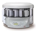
The Leica TP1020 Benchtop autoembedder is a benchtop tissue processor designed to provide gentle dehydration, clearing and infiltration of various tissues with paraffin wax. The TP1020 is completely programmable, allowing tissue-specific programs to be developed. A programmable, optional vacuum setting is available to improve infiltration.
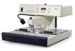
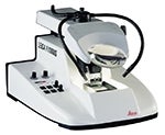
The Leica EG1150H is a heated paraffin dispensing module with a large heated work surface and storage areas for both cassettes and molds. The embedding module aids in obtaining proper orientation of tissues and final embedding in blocks of paraffin for sectioning.
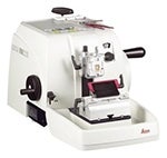
The Leica RM2235 Rotary Microtome is designed for precision manual sectioning of paraffin embedded tissues. The BRC has a knife holder for low profile blades, and a separate knife holder for high profile blades, used for sectioning harder materials. This microtome has a sectioning range of 1-60 mm.
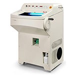
The Leica CM1950 cryostat is available for safe, accurate and fast high quality sectioning. The cryostat can section frozen tissues up to 100 microns in thickness.
The Leica Vibratome VT1000 is allows sectioning of fresh or fixed tissue with or without embedding in support media such as agarose. Speed, frequency, amplitude, and clearance angle are all easily adjustable, and sections can be cut up to 999 microns.
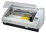
The Leica Autostainer XL provides a gentle method of staining multiple slides. This system is currently set up to quickly and easily stain tissues with hematoxylin and eosin and/or to simply dewax slides cut from paraffin blocks. The processor has the capability to automate multiple staining protocols, with up to 15 programs of 25 steps each.
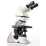
The Leica DM 1000 LED transmitted light microscope features long-life LED illumination that provides near daylight, bright illumination with constant color temperature. This is an extremely versatile microscope system, and can be optionally equipped for dark field, phase contrast, polarization contrast, and fluorescence. The microscope is fitted with a third generation SPOT TM RT3 Slider camera, which allows the user to capture both true color imaging and high sensitivity monochrome images.
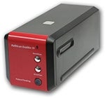
The PathScan Enabler IV Slide Scanner is designed to allow the scanning of an entire whole-mounted histology sample attached to a standard 1 x 3 inch cover-slipped glass slide. This allows for the production of low power, large field of view, high resolution imaging. Pathscan produces image files that can be saved in a variety of formats including TIFF and JPEG.