Fred Christensen, Micah Drew, Samantha Krammer, Dr. Tyler Brown

Introduction
Musculoskeletal injury, particularly in the lower limb, commonly occurs military training, where personnel routinely carry body borne loads between 20 kg and 40 kg [1].
These injuries are attributed to the lower limb biomechanical adaptations during training-related tasks, such as prolonged walking, that result from the heavy body borne loads personnel are required to carry. Yet, it is currently unknown if body borne load or duration of walking lead to intra-limb asymmetries in lower limb biomechanics and increased injury risk during routine training tasks.
Purpose
This study sought to determine the degree to which military body borne load or duration of walking changed lower limb biomechanics.
Methods
Participants
15 (10 male / 5 female) recreationally active adults.
Conditions
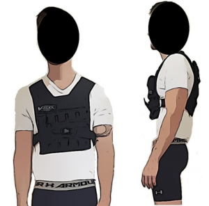
Three body borne loads (unloaded: 0 kg, 15 and 30 kg loads) (Fig. 1).
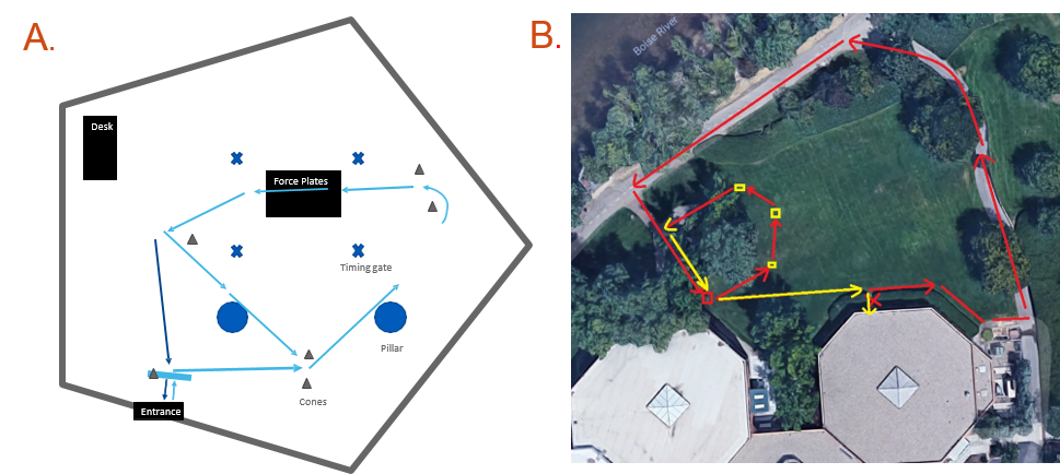
Load Carriage Task
Required participants walk 1.3 m/s for 60 min over-ground with each body borne load (Fig. 2).
Biomechanical Data
Synchronous GRF data and 3D marker trajectories were collected using eight high-speed optical cameras (MXF20, Vicon Motion Systems LTD, Oxford, UK) and a single force platform (OR6, AMTI, Watertown, MA) for three walk trials (1.3 ± 5% m/s) at minutes 0, 30 and 60 during the load carriage task.
Biomechanical Analysis
The marker trajectories were filtered through a 4th order low pass Butterworth filter (12 Hz) and then processed to obtain 3D lower limb (hip, knee, ankle) joint rotations using Visual 3D (C-Motion, Rockville, MD).
Trend Symmetry
Custom written Matlab code calculated trend symmetry according to [2]. Specifically, raw trend symmetry, range amplitude, range offset, and phase shift were calculated for dominant limb (hip, knee and ankle) joint rotations in all three planes.
Statistical Analysis
Trend symmetry parameters were calculated for hip, knee, ankle motions and submitted to a linear model with time (0, 30, 60 minutes) and load (0, 15, 30 kg) as fixed effects. Bonferroni procedure was used for multiple comparisons. Alpha was p < 0.05.
Results
Table 1. Trend symmetry parameters for each time point (minute 0 through 60) for each lower limb joint and plane of motion
| Lower Limb Joint Plane of Motion | Raw Symmetry | Range Amplitude | Range Offset | Phase Shift |
|---|---|---|---|---|
| Hip Flexion | (0.994 - 0.999) | (0.967 – 0.977) | (-0.453 – 0.785) | (0.000 - 0.026) |
| Hip Abduction | (0.914 – 0.946) | (1.014 – 1.032) | (-0.301 – 0.424) | - |
| Hip Rotation | (0.823 – 0.45) | (0.926 - 0.985) | (1.429 - 2.139) | - |
| Knee Flexion | (0.973 - 0.978) | (0.920 – 0.955) | (0.444 - 0.782) | (0.000 – 0.096) |
| Knee Abduction | (0.805 – 0.824) | (0.888 - 0.952) | (-0.046 - -0.590) | - |
| Knee Rotation | (0.876 – 0.894) | (1.023 - 1.102) | (0.759 – 1.475) | - |
| Ankle Flexion | (0.947 – 0.973) | (0.911 - 0.935) | (-1.347 - -1.440) | (-0.230 – 0.043) |
| Ankle Abduction | (0.917 – 0.927) | (1.047 – 1.052) | -0.542 – 0.214) | - |
| Ankle Rotation | (0.958 - 0.976) | (1.039 – 1.081) | (-2.119 - -0.922) | - |
Raw Trend Symmetry
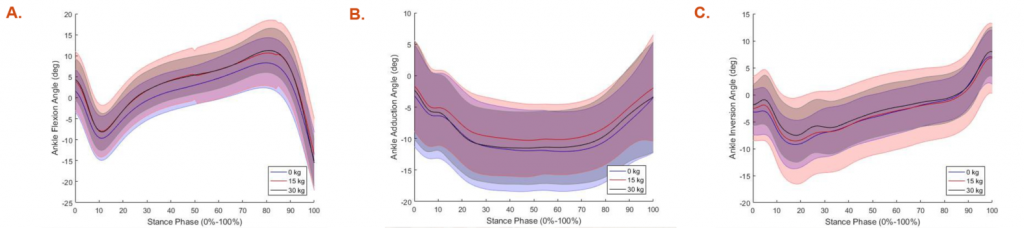
Time had a significant effect on raw symmetry of hip adduction (p=0.035) and ankle internal rotation (p<0.001) (Table 1 and Fig. 3).
Range Offset
Load impacted the range offset of ankle inversion motion (p=0.021), but no hip or knee motion (p>0.05) (Table 1 and Fig. 3).
Range Amplitude
Load effected range amplitude of knee (p=0.049) and ankle flexion (p=0.030) motions, and time effected hip internal rotation motion (p=0.001) range amplitude (Table 1 and Fig. 3).
Phase Shift
Neither load, nor time produced a phase offset in hip, knee, or ankle motion (p>0.05).
Conclusion
Body borne load and duration of walking may elevate injury risk by altering lower limb biomechanics and range of motion during routine military training exercises.
References
- Kaufman, K et al. (2000) AJPM Military Injuries, 54-63.
- Crenshaw, J. et al. (2006) Method for Analyzing Joint Symmetry.
Acknowledgements
MW CTR-IN (subaward # GR07323) providing funding for this work.
Additional Information
Download Trend Symmetry Poster (PDF)
For questions or comments about this research, contact Fred Christensen at fredchristensen@u.boisestate.edu.