Histology

Welcome to the BRC Histology Facility!
Our mission is to provide access to a wide range of histological services for Boise State investigators, external academic investigators and industrial partners. We can provide direct services, training and/or access to equipment for embedding, sectioning and staining of various tissue specimens. We are a fee-for-service center, and use of our facility requires users to establish an ilabs account. To establish an account, or to ask questions regarding our equipment and/or services, please contact:
Cindy Keller-Peck, PhD
Histology Facility Manager
Biomolecular Research Center
1910 University Drive
Boise, ID 83725-1511
Phone: (208) 426-2254
Email: CKPeck@boisestate.edu
Services
Paraffin Embedding
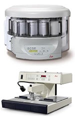
The BRC uses a Leica TP1020 benchtop tissue processor to dehydrate, clear and infiltrate tissues with paraffin. Embedding is done with the aid of a Leica Tissue Embedding Center. Completed blocks will fit in most standard microtome specimen clamps. We currently do not offer training for this equipment.
Paraffin Sectioning
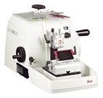
The BRC uses a Leica RM2235 manual rotary microtome to section blocks of paraffin embedded tissues. Most sections are cut at 8 mm, but the microtome has a sectioning range of 1-60 mm in thickness. We can section tissue regardless of whether it was embedded in our facilities. The microtome is available for independent use. We will train users who wish to learn about the technique or would like to section their own tissues.
Frozen Sectioning
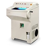
Tissues for frozen sectioning can be prepared according to investigator needs. Fresh tissues are generally snap frozen in OCT using liquid nitrogen/isopentane. Fixed, cryoprotected tissues may be frozen using liquid nitrogen or dry ice. The BRC currently uses a Leica CM1950 clinical cryostat for sectioning frozen tissues. It has a sectioning range of 1-100 mm. The cryostat is available for independent use. If requested, we will train users to freeze and section their own tissues.
Vibratome Sectioning
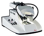
Thick sections of tissues are cut on the Leica VT1000 vibratome. It has a sectioning range of 10-999 mm. Additional adjustable parameters include blade clearance angle, frequency, amplitude and sectioning speed. Tissues with a high degree of structural integrity may be sectioned without a support medium. In instances where a tissue requires support, tissue may be embedded in low melting point agarose. The vibratome is available for independent use. If requested, we will train users to embed and section their own tissues.
Staining
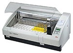
The histology facility currently offers staining with the following histological dyes: Hematoxylin & Eosin, Alcian Blue, and Sirius Red. New dyes and immunohistochemistry services are being added as needed, please email the facility for special requests. We are not currently training investigators to use the Leica Autostainer. However, we will train interested parties in staining techniques to be used back in their individual laboratories.
Rate Table
| Service | Equipment | Training Available |
|---|---|---|
| Paraffin Embedding | Lecia TP1020/Leica EG1150 embedding equipment | No* |
| Paraffin Sectioning | Lecia RM2235 Microtome | Yes |
| Frozen Sectioning | Leica CM1950 Cryostat | Yes |
| Vibratome Sectioning | Lecia Vibratome VT1000 | Yes |
| Staining | Leica Autostainer XL | No* |
For rate information please see Histology BRC Recharge Rates
*May be available by special request.
Sample Submission
Users who wish to submit samples for processing must first obtain an iLabs account. A one-time registration is required to place service requests or reserve equipment. Please visit the iLabs registration page. For questions on using iLabs to set up histology services, please contact the Histology Facility Manager.
Once an iLabs account is established, and a service request or equipment reservation is made, tissue should be delivered to the Histology Lab in room 211 of the Math building. The tissue should be accompanied by a completed copy of the Histology Submission Form. This form may also be filled out and emailed to CKPeck@boisestate.edu.
Tissue Preparation: Tissues are most commonly submitted to the BRC in 10% neutral buffered formalin (NBF). However, tissues are accepted in other fixatives provided the researcher feels it is the most appropriate way to maintain the molecular and macromolecular aspects under investigation.