Fabio Halla, Emmie Jennings, Akina Fujimoto, Stephanie Tuft, Dr. Julia Oxford
Abstract
Dexamethasone, a corticosteroid that inhibits the release of substances in the body that cause inflammation, is commonly used for the treatment of arthritis. However, glucocorticoid-induced osteoporosis is a side effect that commonly occurs after dexamethasone treatment. One of the mechanisms in which glucocorticoids is thought to suppress bone formation is through their effect on the wnt/β-catenin signaling pathway. The wnt/β-catenin pathway is essential in the formation of new osteoblasts and the prevention of current osteoblast apoptosis. However, treatment with dexamethasone is thought to destabilize and inhibit nuclear translocation of β-catenin, decreasing the survival of osteoblasts.1 By using precursor mouse osteoblast MC3T3 cells, antisense collagen 11 is analyzed as a potential treatment to reverse the detrimental effects of dexamethasone on the wnt signaling pathway. MC3T3 cells are exposed to dexamethasone in vitro, then aspects of the wnt pathway and collagen system are studied in depth.
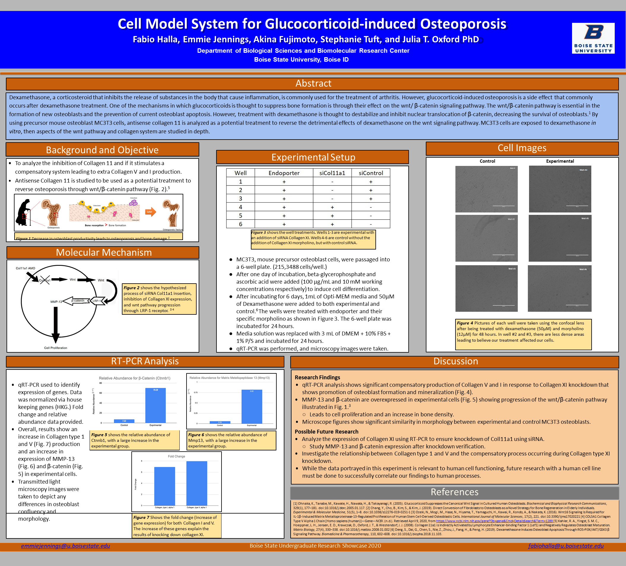
Background and Objective
- To analyze the inhibition of Collagen 11 and if it stimulates a compensatory system leading to extra Collagen V and I production.
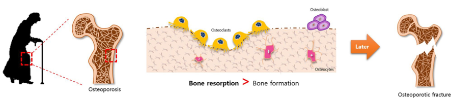
- Antisense Collagen 11 is studied to be used as a potential treatment to reverse osteoporosis through wnt/β-catenin pathway (Fig. 2).(Reference 5)
Molecular Mechanism
Experimental Setup
- MC3T3, mouse precursor osteoblast cells, were passaged into a 6-well plate. (215,3488 cells/well.)
- After one day of incubation, beta-glycerophosphate and ascorbic acid were added (100 µg/mL and 10 mM working concentrations respectively) to induce cell differentiation.
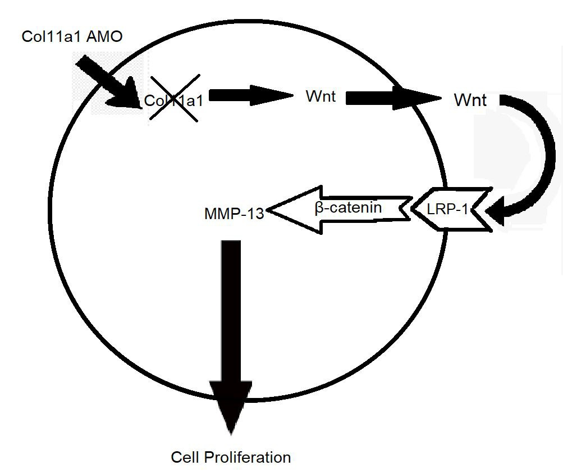
- After incubating for 6 days, 1mL of Opti-MEM media and 50µM of Dexamethasone were added to both experimental and control.6 The wells were treated with endoporter and their specific morpholino as shown in Figure 3. The 6-well plate was incubated for 24 hours.
- Media solution was replaced with 3 mL of DMEM + 10% FBS + 1% P/S and incubated for 24 hours.
- qRT-PCR was performed, and microscopy images were taken.
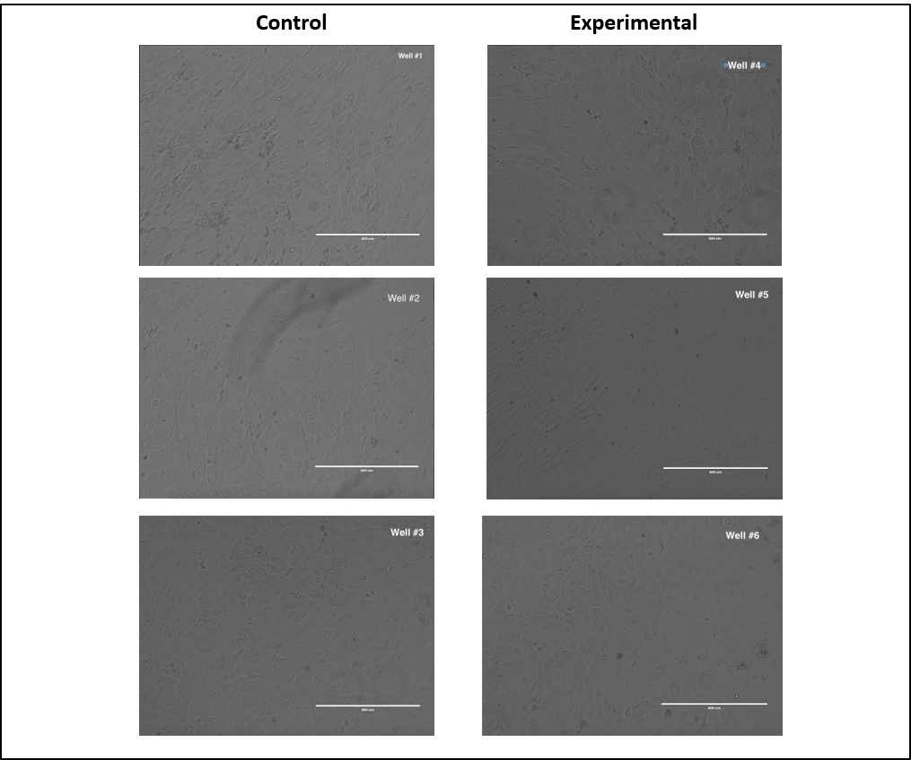
Cell Images
RT-PCR Analysis
- qRT-PCR used to identify expression of genes. Data was normalized via house keeping genes (HKG.) Fold change and relative abundance data provided.
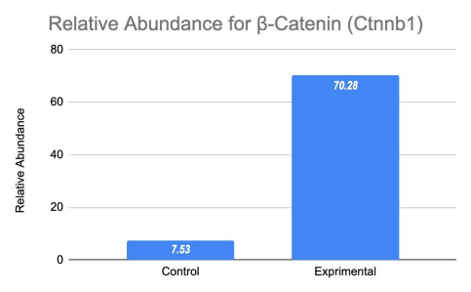
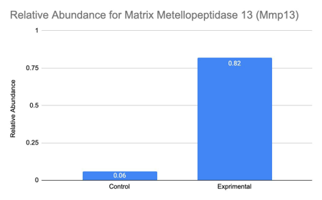
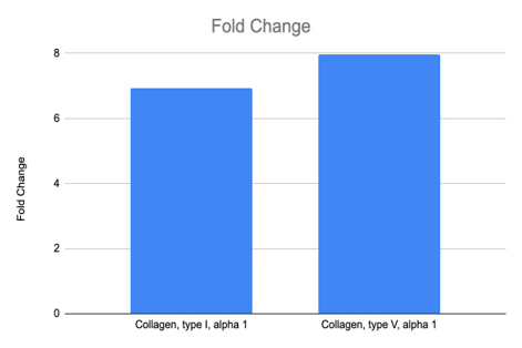
- Overall, results show an increase in Collagen type 1 and V (Fig. 7) production and an increase in expression of MMP-13 (Fig. 6) and β-catenin (Fig.5) in experimental cells.
- Transmitted light microscopy images were taken to depict any differences in osteoblast confluency and morphology.
Discussion
Research Findings
- qRT-PCR analysis shows significant compensatory production of Collagen V and I in response to Collagen XI knockdown that shows promotion of osteoblast formation and mineralization (Fig. 4).
- MMP-13 and β-catenin are overexpressed in experimental cells (Fig. 5) showing progression of the wnt/β-catenin pathway illustrated in Fig. 1. (Reference 3)
- Leads to cell proliferation and an increase in bone density.
- Microscope figures show significant similarity in morphology between experimental and control MC3T3 osteoblasts.
Possible Future Research
- Analyze the expression of Collagen XI using RT-PCR to ensure knockdown of Coll11a1 using siRNA.
- Study MMP-13 and β-catenin expression after knockdown verification.
- Investigate the relationship between Collagen type 1 and V and the compensatory process occurring during Collagen type XI knockdown.
- While the data portrayed in this experiment is relevant to human cell functioning, future research with a human cell line must be done to successfully correlate our findings to human processes.
References
- Ohnaka, K., Tanabe, M., Kawate, H., Nawata, H., & Takayanagi, R. (2005). Glucocorticoid Suppresses the Canonical Wnt Signal in Cultured Human Osteoblasts. Biochemical and Biophysical Research Communications, 329(1), 177–181. doi:10.1016/j.bbrc.2005.01.117.
- Chang, Y., Cho, B., Kim, S., & Kim, J. (2019). Direct Conversion of Fibroblasts to Osteoblasts as a Novel Strategy for Bone Regeneration in Elderly Individuals.
Experimental & Molecular Medicine, 51(5), 1–8. doi:10.1038/s12276-019-0251-1 - Ozeki, N., Mogi, M., Hase, N., Hiyama, T., Yamaguchi, H., Kawai, R., Kondo, A., & Nakata, K. (2016). Wnt16 Signaling Is Required for IL-1β-Induced Matrix Metalloproteinase-13-Regulated Proliferation of Human Stem Cell-Derived Osteoblastic Cells. International Journal of Molecular Sciences, 17(2), 221. doi:10.3390/ijms17020221
- COL5A1 Collagen Type V Alpha 1 Chain [Homo sapiens (human)]—Gene—NCBI. (n.d.). Retrieved April 9, 2020, from https://www.ncbi.nlm.nih.gov/gene?Db=gene&Cmd=DetailsSearch&Term=1289
- Kahler, R. A., Yingst, S. M. C., Hoeppner, L. H., Jensen, E. D., Krawczak, D., Oxford, J. T., & Westendorf, J. J. (2008). Collagen 11a1 is Indirectly Activated by Lymphocyte Enhancer-binding Factor 1 (Lef1) and Negatively Regulates Osteoblast Maturation. Matrix Biology, 27(4), 330–338. doi:10.1016/j.matbio.2008.01.002
- Deng, S., Dai, G., Chen, S., Nie, Z., Zhou, J., Fang, H., & Peng, H. (2019). Dexamethasone Induces Osteoblast Apoptosis Through ROS-PI3K/AKT/GSK3βSignaling Pathway. Biomedicine & Pharmacotherapy, 110, 602–608. doi:10.1016/j.biopha.2018.11.103.
Additional Information
For questions or comments about this research, contact Fabio Halla at fabiohalla@u.boisestate.edu.