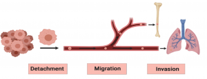
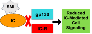
The cell signaling pathways activated by a specific IC binding to the cell membrane receptors of cancer cells, initiating early stages of metastasis.
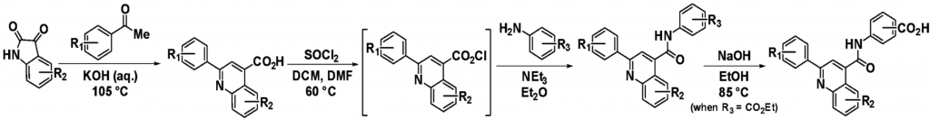
General Synthetic Scheme for IC-SMI-26 Analogs
Synthesis begins with a Pfitzinger reaction, followed by an acid chloride formation/amidation sequence to form the aryl amid intermediate, and lastly, a subsequent hydrolysis of the ethyl ester
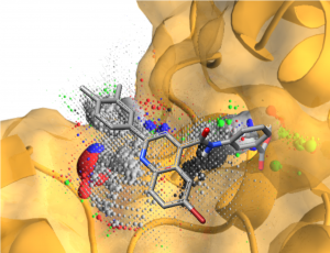
Computational Modeling for SMI-26 Binding Interactions with the IC
(A) SMI-26 overlaid on a computational binding probability density map of the IC(B) 3D model representing important amino acid contributions to SMI-26 binding.
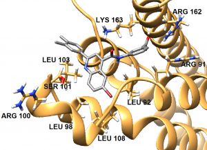
Enzyme-Linked Immunosorbent Assay (ELISA) of SMI-26 Analogs
Bar Graph of the Relative pSTAT3 Expression of SMI 26 and Analogs in T47D Breast Cancer cells. The Y-axis is the relative pSTAT3 expression of the different treatment of SMI Analogs (x-axis). The two controls are the No Treatment (no IC) with PSTAT3 level around 0.2 and the IC PSTAT3 level normalized to 1. In the middle of the graph is a struture of SMI-26 analogs with labeled aryl groups. Aryl group 1 contains a subsituted benzene ring and is labled green at the top left. Aryl group 2 is a quinoline core structure colored red. Aryl group 3 is a benzamide group labled blue at the top right.Six SMI-26 analogs made with aryl group 1 (colored green on compound structure in graph) changed with the following groups: benzene ring with no substituents, benzene ring with a chlorine atom in the para position, benzene ring with a methyl group in the para position, benzene ring with two chlorine atoms in the para and meta positions, benzene ring with a chlorine atom in the meta position, and a benzene ring with an ether group in the para position.Six SMI-26 analogs made with the quinoline base structure, aryl group 2 (colored red on compound structure in graph) changed with the following groups: no substituents, chlorine atom in position 8, two chlorine atoms in positions 8 and 7, methyl group in position 6, fluorine atom in position 6, bromine atom in position 7.12 SMI-26 analogs made with benzylamide group 3 (colored blue on compound structure in graph) changed with the following groups on the benzene ring: chlorine atom in the meta position, tri-fluoro (3 fluorine atoms bonded to a single carbon atom) group in the para position, a benzyl group, 3-methylpyridine group, benzyl-tri-fluoro group, and a carboxylic acid group in the para position
Analogs were systematically optimized by changing one of the three aryl (Ar) moieties at a time followed by subsequent ELISA tests for indirect binding affinity
Lower pSTAT3 expression is indicative of lower cell signaling activity
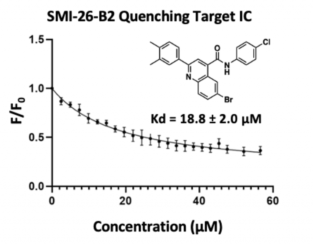
Fluorescence Quenching Assay
A Graph of the Fluorescence Quenching Assay SMI-26-B2 with the target IC. The y-axis is the measured normalized fluorescence intensity at each titration point divided by the fluorescence intensity at the 0 point (F/F0) for given concentrations of SMI in micromolar. SMI was added to IC sample in increments of 5 micromolar. A Concave, exponential decay curve is drawn to best fit the data points. The reported Kd value is 18.8 plus or minus 2.0 (at the 95% confidence level) micromolar.
Assay quantitatively measures direct SMI binding to target IC
Lower Kd is indicative of stronger binding
Errors bars are reported at the 95% Confidence Level
Conclusion
Incorporation of halogen substituents in aryl group 2 and a strong electron withdrawing group in the para position of aryl group 3 show increased binding affinity
It is predicted hydrophobic interactions play a crucial role for binding in aryl group 1
Fluorescence experiment indicates direct binding and inhibition of target IC
Future Work
Measure the binding affinity of all SMI-26 analogs to the IC by fluorescence quenching assays
Complete full characterization of synthesized SMI-26 analogs
Further optimize analogs for superiorIC inhibitionandimproved drug-likeness
Acknowledgements
The Institutional Development Awards (IDeA) from the National Institute of General Medical Sciences of the National Institutes of Health under Grant Nos. P20GM103408 and P20GM109095, The Biomolecular Research Center at Boise State with funding from the National Science Foundation, Grant Nos. 0619793 and 0923535, the MJ Murdock Charitable Trust, the Idaho State Board of Education and The METAvivorQuinn Davis Northwest Arkansas METSqueradeFund. The authors appreciatively acknowledge the gracious assistance Dr. Joseph Dumais, Riley Olsen, and Joseph Tuccinardi.