Carey Hammer, TJ Rhodes, Jenna Draper, Susan Pett, Natalie Mourant
Introduction
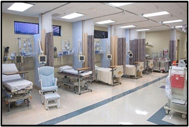
Radiographic technology has increased in popularity among hospitals, doctors’ offices, and clinics. With this increase in accessibility, the potential for relaxed standards in radiation protection have heightened. The use of portable radiographic machines have made it possible to conduct exams in many convenient locations, notably areas lacking in adequate structural shielding. In many medical facilities, recovering patients are separated by a thin curtain or non-shielded barrier. This is a risk to patients and bystanders potentially receiving unnecessary dose. The purpose of this experiment is to determine if there is significant radiation penetrating through non-shielded barriers.
Methods
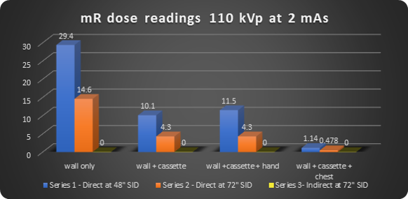
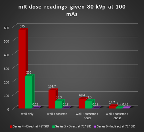
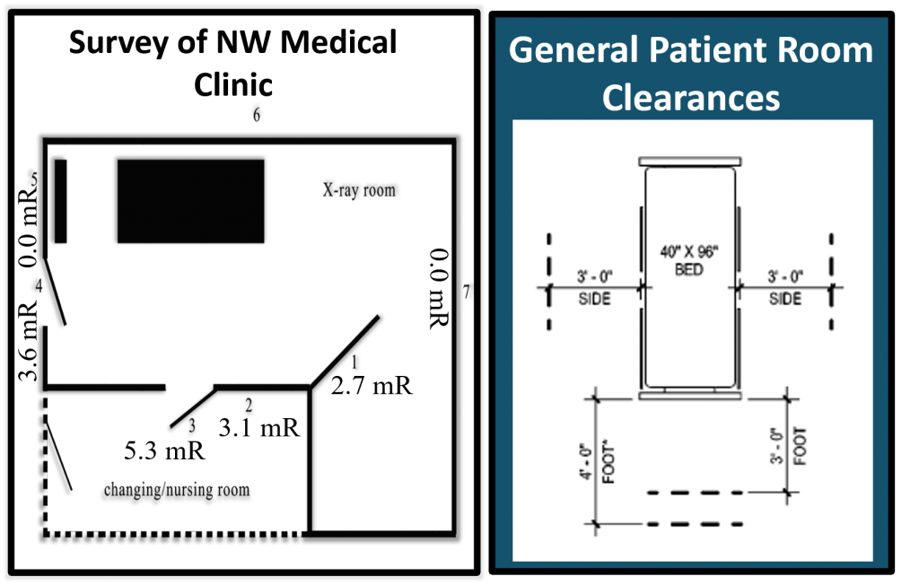
Data was collected from three sources for this experiment. Radiation dosage readings were obtained for the first set of data from a portable GE radiographic machine with only the wall as a barrier, located at a college health science building . A dosimeter was placed in adjoining room and attached to the wall at the same height as the central beam and measurements take (see graph). Additional exposures were performed through the wall at varying distances with either a hand, or thoracic phantom. The second set of data was completed on June 14, 2019 by a team of physicists at a northwest medical clinic site. Dosimeters were placed outside each of the four walls, the entryway door, the changing room door, and the control booth window. Measurements were then recorded. Dosimeter readings were obtained for the third set of data at the same college health science building and utilizing the same portable machine at three and six feet to mimic general patient recovery room clearances.
Results
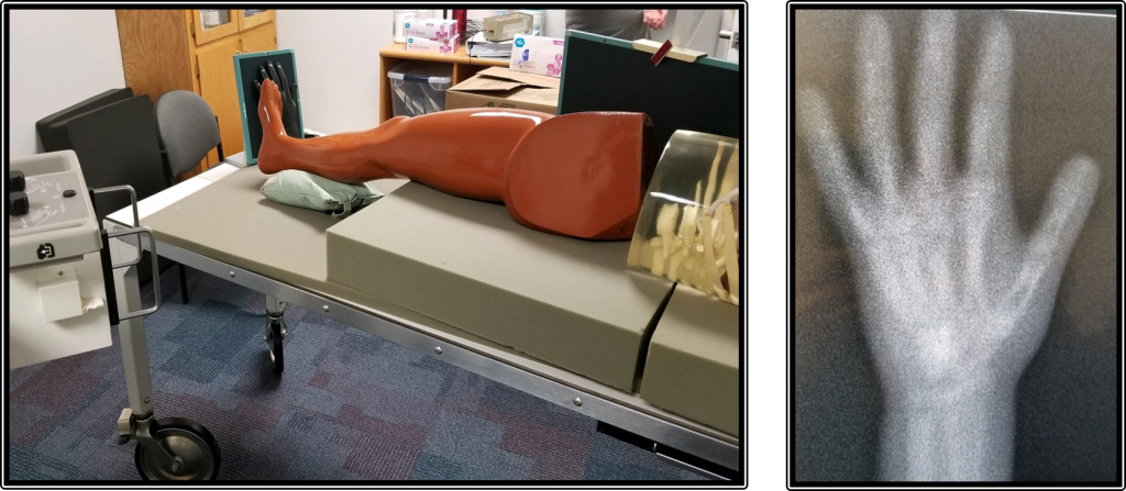
Further studies were conducted to obtain radiation exposure readings at three feet and six feet from the patient to mimic the general patient room clearances with curtains as the barrier between rooms. The three-foot measurement was based on the amount of radiation dose a guest of the patient may receive sitting in a chair next to the adjacent curtain. The six-foot clearance was to measure the amount of ionizing radiation the patient in the next room may be unwittingly exposed to. The below left image demonstrates the experiment set up. The below right image is the indirect exposure to the hand phantom placed in front of a CR cassette at the foot of the “patient’s” bed, three feet from the area of collimation.
Dosimeter Readings at 3 feet – kVp 80 @ mAs 40
| Location | Reading |
|---|---|
| Dosimeter behind plate – tight collimation | 1.074 mR |
| Dosimeter behind plate – open collimation | 4.76 mR |
| Dosimeter at a 45o angle from plate | 0.943 mR |
| Hand phantom on CR plate visible | Yes – placed at foot |
Dosimeter Readings at 6 feet – kVp 80 @ mAs 40
| Location | Reading |
|---|---|
| Dosimeter behind plate – tight collimation, hand phantom at feet | 0.424 mR S# 2704 |
| Dosimeter behind plate – open collimation, hand phantom at 6 feet | 0.614 mR S # on hand phantom 1959 |
| Hand phantom at 6 feet – tight collimation | S# 1592 |
The charts seen directly above from the third set of data depict the dosimeter readings at three and six feet from the edge of the bed. The readings at six feet also include the S# from the CR reader which has an indirect relationship to dose. Based on the CR reader used, the typical diagnostic image range is between 100-400. The readings indicate low radiation exposure to the hand phantom; however, an image was formed in all three exposures due to indirect scatter radiation penetrating through the hand phantom and exposing the CR plate.
Occupational workers – State and national standards comparison with NW clinic
| Idaho | NCRP | Clinic |
|---|---|---|
| 2 mR per hour | 2.5 mR per hour (whole body), 150 mSv lens of eye, Lowered to 50 mGy = 5700 mR per yr | #1) 60 mR per hour (eyes) |
Non-controlled (unrestricted) areas – State and national standards comparison with NW clinic
| Idaho | NCRP | Clinic |
|---|---|---|
| 2 mR per week | 2.2 mR per week | #2) 3.1 mR per week #3) 5.3 mR per week #4) 3.6 mR per week |
The results of this study determined there is a significant amount of x-ray penetration through both unleaded barriers. In many instances, the dose of scatter radiation exceeded the maximum standards for x-ray exposure in unrestricted areas. This experiment indicated the necessity of collimating correctly, shielding, and maintaining distance from the radiation source. The NCRP recommends a distance of six feet or more for patients in adjoining rooms. Radiographers should be mindful of family members that may be obscured by curtains to ensure proper distance. Precautions must be taken by medical facilities to demonstrate ALARA practices that protect both occupational workers and patients.
Future Research
This research was limited to three different locations and methods. Further research could include data from multiple locations and effects of increased mAs to determine if the general guidelines of an eight-foot distance between beds is sufficient for recovery rooms.
References
- Idaho Department of Health and Welfare. (n.d.). Idaho Radiation Control Rules (16.02.27). Retrieved from https://adminrules.idaho.gov/rules/2000/16/0227.pdf
- Nkubli, F.B., Nzotta, C.C., Nwobi, N.I., Moi, S.A., Adejoh, T, Luntsi, G., Dlama, J., Shem, L.S. (2017). A survey of structural design of diagnostic x-ray imaging facilities and compliance to shielding design goals in limited resource setting. The Journal of Global Radiology, 3(1): Article 6. doi:10.7191/jgr.1017.1041
- Carroll, Q. B. (2018). Radiography in the digital age (3rd ed.) Springfield, IL: Charles C. Thomas
Additional Information
For questions or comments about this research, contact Carey Hammer at careyhammer@u.boisestate.edu.